

Cone beam imaging changes how your dentist sees your teeth, bones, and joints. Traditional X‑rays give flat pictures. Cone beam imaging gives a clear 3D view. You get sharper detail. Your dentist gets stronger facts. This helps find problems early. It also helps plan treatment that fits your mouth. You spend less time guessing and more time healing. If you see a general dentist tampa fl, cone beam imaging can show hidden infections, nerve paths, and jaw issues before they turn serious. You and your dentist can talk through clear images together. You can see what is wrong and what comes next. This brings calm during stressful visits. It also cuts the risk of surprises during surgery or root canals. With better pictures, your dentist can plan with confidence, protect healthy tissue, and choose the safest path for your care.
What Cone Beam Imaging Actually Is
Table Contents
- What Cone Beam Imaging Actually Is
- How It Compares To Traditional Dental X‑Rays
- Conditions Cone Beam Imaging Helps Find
- Why It Matters For Treatment Planning
- 1. Safer implant placement
- 2. Root canal success
- 3. Jaw joint and bite problems
- What The Scan Feels Like For You
- Radiation And Safety
- How Cone Beam Imaging Helps Your Family
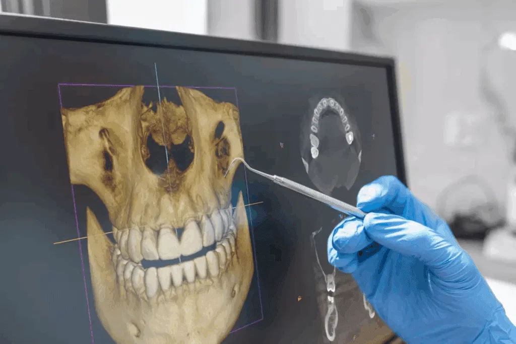
Cone beam imaging is a special type of dental CT scan. The machine moves in a circle around your head. It collects hundreds of small images. A computer turns these images into a 3D model of your teeth, jaw, and face.
You do not feel pain. You stand or sit still for less than a minute. The scan is quiet. You keep your clothes on. You bite on a small piece of plastic to help keep your head steady. Then the scan ends.
This 3D model lets your dentist look from any angle. Your dentist can zoom in on one tooth, a joint, or a nerve. That level of detail is not possible with basic X‑rays.
How It Compares To Traditional Dental X‑Rays
Traditional bitewing and panoramic X‑rays still help. They show cavities, bone levels, and basic tooth shapes. Yet they flatten your mouth into one layer. This can hide trouble spots.
Cone beam imaging adds depth. It shows the width, height, and depth of bone and teeth. It also shows how structures relate to each other. That difference matters when you need precise care.
| Feature | Traditional Dental X‑rays | Cone Beam Imaging
|
|---|---|---|
| Type of image | 2D flat picture | 3D model of teeth and jaws |
| Detail of bone and roots | Basic shapes | Clear root shape, bone height, bone width |
| View of nerves and sinuses | Hard to see | Shows nerve paths and sinus spaces |
| Use for surgery planning | Limited | Strong support for implants and jaw surgery |
| Radiation dose | Low | Low to moderate but focused on a small area |
| Patient position | Sitting or standing still for seconds | Sitting or standing still for under a minute |
The American Dental Association states that dentists should use the lowest radiation needed to answer clear questions. Cone beam imaging follows that rule when your dentist uses it only when needed and sets the smallest scan size that still gives answers.
Conditions Cone Beam Imaging Helps Find
Cone beam imaging helps your dentist see problems that stay hidden on standard X‑rays. Common uses include three groups.
- Teeth and roots. Cracks in roots. Extra roots. Impacted teeth that did not come in.
- Bone and joints. Bone loss from gum disease. Jaw joint changes that cause pain or popping.
- Other structures. Cysts, small growths, or sinus issues that press on teeth.
When your dentist can see the full shape of a tooth and the bone around it, the treatment plan changes. A tooth that looks hopeless on a flat X‑ray may be worth saving when seen in 3D. The opposite can also be true. That honesty protects you from repeated work and long pain.
Why It Matters For Treatment Planning
Better images lead to clearer plans. This helps in three main ways.
1. Safer implant placement
Dental implants need enough bone in height and width. The implant must also avoid nerves and the sinus. Cone beam imaging measures bone in all directions. Your dentist can see the exact spot where the implant should go. Your dentist can also see if you need bone grafting first.
2. Root canal success
Root canals fail when a canal is missed or a crack goes unseen. Cone beam imaging shows extra canals, narrow canals, and small fractures. It also shows infection in the bone that a flat X‑ray can miss. This raises the chance that one root canal is enough.
3. Jaw joint and bite problems
Jaw joint pain and bite issues confuse many people. Cone beam imaging shows the joint surfaces and the way the jawbone sits in the joint. It also shows how upper and lower teeth meet. This helps your dentist plan bite guards, braces, or other care with more accuracy.
What The Scan Feels Like For You
You stand or sit. The assistant lines up the machine. You may bite on a small tab. The arm moves around your head in a circle. You breathe as normal. The scan ends in less than a minute.
There are no needles. There is no contact with your skin other than the bite tab or a small support. Children and adults can both complete the scan without trouble. If you feel nervous, tell the team. They can show the steps first, so you feel in control.
Radiation And Safety
Radiation always raises concern for families. That concern is reasonable. Cone beam scans use more radiation than a single small dental X‑ray. Yet they often use less than a full medical CT of the head.
The U.S. Food and Drug Administration gives guidance on safe dental imaging. Dentists are trained to follow the ALARA concept. That means “as low as reasonably achievable.” Your dentist will order a cone beam scan only when the extra detail changes your care. Your dentist can also limit the scan to a small region instead of the full head.
You can ask three key questions.
- Why is this scan needed for my case
- Will it change the treatment plan
- Are there other options that use less radiation
Clear answers to these questions should bring peace of mind.
How Cone Beam Imaging Helps Your Family
For you and your family, cone beam imaging means fewer surprises. It can shorten treatment time. It can reduce guesswork. It can help your dentist choose care that fits your mouth, not a standard chart.
With sharper 3D pictures, your dentist can see the full story of your teeth and jaws. That clarity supports early diagnosis, safer surgery, and stronger long-term results. It also supports honest talks. You can see the problem on the screen and understand why a certain path is best.
When you know your dentist is working with complete, clear images, it is easier to sit back in the chair, breathe, and trust the plan.